Loculated Pleural Effusion X Ray : Pleural Effusion Treatment & Management. Check for pleural thickening and pleural effusions. The plain chest radiographic features of pleural effusion are usually characteristic. Us scan they can be identified clearly and it is very complicated.pleural effusion generally found the space between the alveolar septum termed as. 299 370 просмотров 299 тыс. Pleural effusions can loculate as a result of adhesions.
Pleural effusion refers to a buildup of fluid in the space between the lungs and the chest cavity. This patient was known to have pleuritic carcinomatosis. Check for pleural thickening and pleural effusions. Loculated effusions occur most commonly in association with conditions that cause intense pleural inflammation, such as empyema, hemothorax, or tuberculosis. Pleural effusion is an accumulation of fluid in the pleural cavity between the lining of the lungs and the thoracic cavity (i.e., the visceral and parietal pleurae).
Loculated pleural effusion | Radiology Case | Radiopaedia.org from images.radiopaedia.org In the usa approximately 1.5 million people are diagnosed with a pleural effusion each year 2. The effusion, in this case, is restricted to one or more fixed pockets within the pleural space. Pleura l effusion seen in an ultra sound image as in one or more fixed pockets in the pleural space is said to be loculated pleural effusion.in. Pleura is a mesothelial lined sac that envelopes the lungs and comprises of 2 membranous walls i.e. Ct scans show more detail than. This patient was known to have pleuritic carcinomatosis. The patient's history and physical exam may indicate a presumptive. Occasionally, a focal intrafissural fluid collection may look like a lung mass.
Us scan they can be identified clearly and it is very complicated.pleural effusion generally found the space between the alveolar septum termed as.
Pleura is a mesothelial lined sac that envelopes the lungs and comprises of 2 membranous walls i.e. It allows distinction between free and loculated fluid showing its extent and localization. no change in position of effusion withchange in position of chest. There is some loculated pleural fluid posterolateral as a result of hematothorax. Conventional radiography is usually the first step in the detection of a pleural effusion. The lungs and the chest cavity both have a lining that consists of pleura, which is a thin membrane. It can result from pneumonia and many other conditions. Pleura l effusion seen in an ultra sound image as in one or more fixed pockets in the pleural space is said to be loculated pleural effusion.in. This patient was known to have pleuritic carcinomatosis. In healthy lungs, these membranes ensure that a. Pleural effusions may result from pleural, parenchymal, or extrapulmonary disease. Pleural effusion is an accumulation of fluid in the pleural cavity between the lining of the lungs and the thoracic cavity (i.e., the visceral and parietal pleurae). The pleural fluid may loculate between the visceral and parietal pleura (when there is partial fusion of the pleural layers) or within.
The annual incidence of pleural effusion in the developed world has been estimated at 320 per 100,000 population per year 1. Pleural effusions can loculate as a result of adhesions. A pleural effusion is accumulation of excessive fluid in the pleural space, the potential space that surrounds each lung. Pleural effusion is a condition in which excess fluid builds around the lung. Check for pleural thickening and pleural effusions.
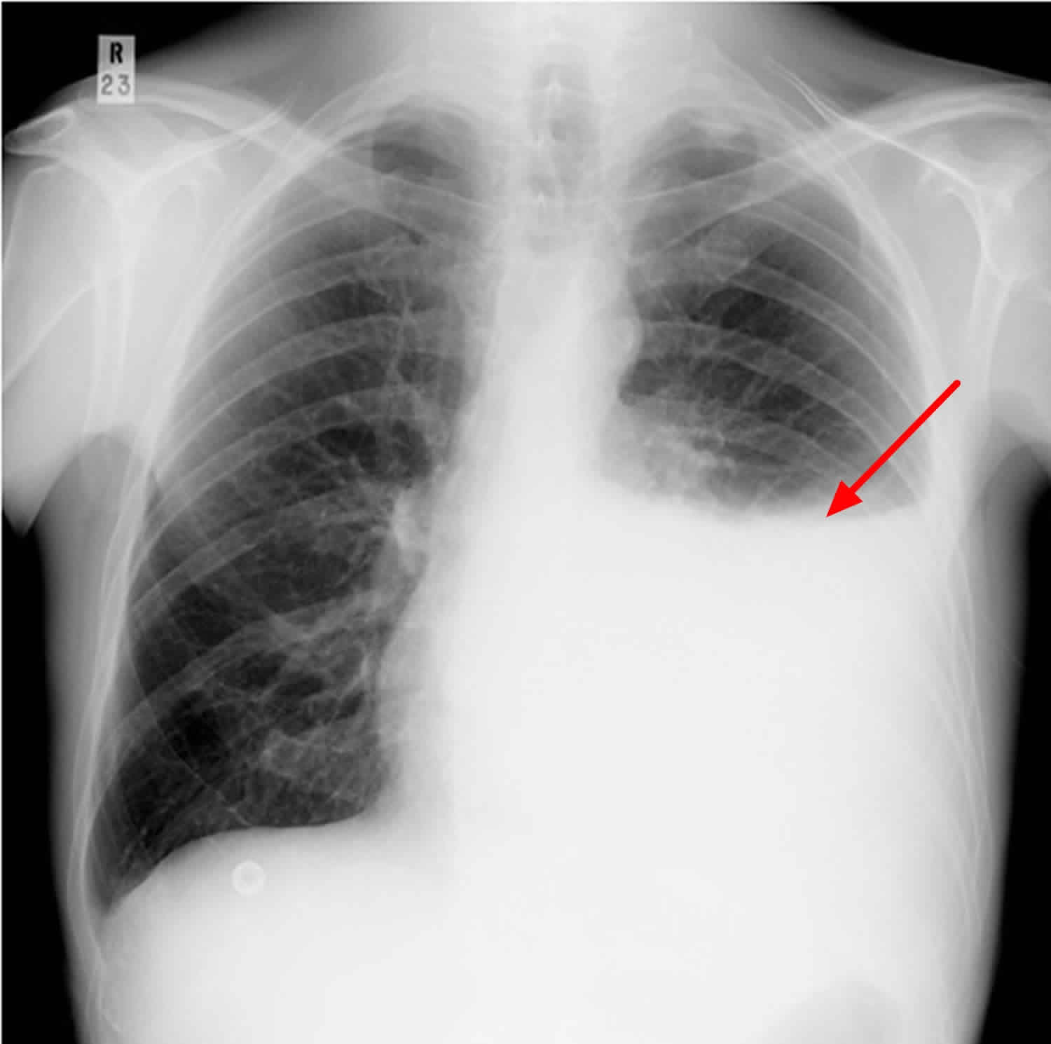
Loculated pleural effusion | Radiology Case | Radiopaedia.org from images.radiopaedia.org A pleural effusion is an abnormal collection of fluid within the pleural space. Conventional radiography is usually the first step in the detection of a pleural effusion. It can result from pneumonia and many other conditions. The left lung is almost. The patient's history and physical exam may indicate a presumptive. The left lower zone is uniformly white. Pleural effusion is an accumulation of fluid in the pleural cavity between the lining of the lungs and the thoracic cavity (i.e., the visceral and parietal pleurae). Pleural effusions can loculate as a result of adhesions.
It can result from pneumonia and many other conditions.
When blunting of these costophrenic angles is seen, it is suggestive of. Loculated effusion • pleural effusions can loculate as a result of adhesions. Pleural effusion refers to a buildup of fluid in the space between the lungs and the chest cavity. The annual incidence of pleural effusion in the developed world has been estimated at 320 per 100,000 population per year 1. In the usa approximately 1.5 million people are diagnosed with a pleural effusion each year 2. The left lung is almost. In healthy lungs, these membranes ensure that a. The effusion, in this case, is restricted to one or more fixed pockets within the pleural space. Concave meniscus (horizontal in case of. Conventional radiography is usually the first step in the detection of a pleural effusion. Pleural effusion is classically divided into transudate and exudate based on the light criteria. 299 370 просмотров 299 тыс. It can result from pneumonia and many other conditions.
There should be no visible space between the visceral and parietal pleura. The left lower zone is uniformly white. Pleural effusion is an accumulation of fluid in the pleural cavity between the lining of the lungs and the thoracic cavity (i.e., the visceral and parietal pleurae). Conventional radiography is usually the first step in the detection of a pleural effusion. This patient was known to have pleuritic carcinomatosis.
Pleural effusion causes, types, symptoms, diagnosis and treatment from healthjade.com If you miss a tension pneumothorax you risk your patient's. Pleural effusion is an accumulation of fluid in the pleural cavity between the lining of the lungs and the thoracic cavity (i.e., the visceral and parietal pleurae). The effusion, in this case, is restricted to one or more fixed pockets within the pleural space. Loss of the costophrenic angle. What procedures and tests diagnose pleural effusions? When blunting of these costophrenic angles is seen, it is suggestive of. Pleural effusions can loculate as a result of adhesions. Ct scans show more detail than.
Ct scan is the most sensitive modality for detection of presence of minimal fluid.
The pleura and pleural spaces are only visible when abnormal. Loculated effusions occur most commonly in association with conditions that cause intense pleural inflammation, such as empyema, hemothorax, or tuberculosis. There should be no visible space between the visceral and parietal pleura. Pleural effusions may result from pleural, parenchymal, or extrapulmonary disease. It allows distinction between free and loculated fluid showing its extent and localization. Ct scans show more detail than. Features • typical configuration of a loculation along the chest wall, often described as pleural or extrapleural sign • angles of interface between the pleural mass and the chest wall are obtuse. What are the pulmonary findings? The left lung is almost. Pleura is a mesothelial lined sac that envelopes the lungs and comprises of 2 membranous walls i.e. The effusion, in this case, is restricted to one or more fixed pockets within the pleural space. When blunting of these costophrenic angles is seen, it is suggestive of. Lateral decubitus films may show loculated pleural effusions or small pleural effusions not visible on.
The pleural fluid may loculate between the visceral and parietal pleura (when there is partial fusion of the pleural layers) or within loculated pleural effusion. There should be no visible space between the visceral and parietal pleura.
Share :
Post a Comment
for "Loculated Pleural Effusion X Ray : Pleural Effusion Treatment & Management"


Post a Comment for "Loculated Pleural Effusion X Ray : Pleural Effusion Treatment & Management"